Screening & Diagnostic Imaging for All Women
We offer specialized care in a technologically advanced facility and foster close partnerships with area physicians and surgeons.
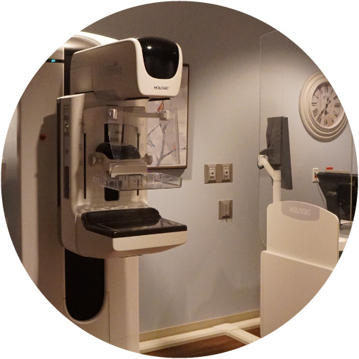
Diagnostic Mammography in Maple Grove, MN
Why is This Exam Done?
Diagnostic mammography is used to evaluate a patient with signs or symptoms such as lumps, nipple discharge, pain, dimpling or puckering of skin. It is also used to further evaluate an area of concern found on a screening mammogram. The images are interpreted (read) by a Radiologist at the time of the exam and the Mammographer will do additional imaging as directed. You will be advised of the results before leaving the facility. You will need an order from your referring physician if you are having anything other than a screening mammogram.
What Will Happen During the Exam?
You will be asked to change into a warm robe undressed from the waist up. A Mammographer (a specially qualified radiologic technologist) will ask you several questions to review your history. These include questions about your family history and any previous breast surgery. Your breast will be placed on a special platform of the mammography unit and the Mammographer will gradually compress your breast, working with you to limit any discomfort. You will be asked to hold very still to limit motion on the images. The Mammographer will reposition you between images and check to ensure the images are well positioned and of good technical quality before discharging you.
How Should I Prepare?
- Do not wear deodorant, talcum powder or lotion under your arms or on your breasts on the day of the exam. These can appear on the mammogram as calcifications (calcium spots) and you may need to return for additional imaging.
- Wear comfortable 2 piece clothing as you will be asked to remove all clothing above the waist and change into a warm robe.
- Always inform the staff if you feel you may be pregnant.
- Allow 15 minutes for a screening mammogram and 30 minutes for a diagnostic mammogram.
- If you have a rash or sore on or beneath your breasts, you should postpone your routine exam until the rash/sore is gone or healed.
Risks you should be aware of:
- About 10% of Screening Mammograms require more testing such as a diagnostic mammogram or ultrasound and most of these result in normal (negative) findings.
- Abnormal findings may require a follow-up short-term mammogram or a biopsy. Most biopsies have normal results (negative) and confirm that no cancer was present.
- Women with dense breast tissue may benefit from supplemental imaging such as Breast MRI.
After your exam:
- Your images will be interpreted (read) by one of our board-certified breast imaging radiologists, Minneapolis Radiology.
- You will be given your results when your exam is complete and the findings will be sent to your healthcare provider.
- In certain circumstances, you may need further diagnostic evaluation by having a breast ultrasound. This does not necessarily indicate an abnormality was found or that your mammogram was not properly obtained, but that additional images are needed to ensure all breast tissue is fully evaluated.