Screening & Diagnostic Imaging for All Women
We offer specialized care in a technologically advanced facility and foster close partnerships with area physicians and surgeons.
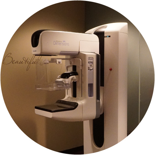
Digital Mammography in Maple Grove, MN
A mammogram is a low-dose x-ray of the breast and surrounding tissues which can effectively detect cancers long before you or your physician might feel any changes. The goal of mammography is early detection of breast cancer, typically through detection of masses and/or micro-calcifications (very small bits of calcium can appear within the soft tissue of your breast).
Need to schedule an appointment? Contact us 24 hours a day using our contact form or call during business hours at (763) 398-6370.
Digital Mammography, also called Full-Filed Digital Mammography (FFDM), is a mammography imaging system where the images are seen on a computer screen and can be saved electronically. The images can then be manipulated by the Radiologist to enhance or magnify an area.
Annual screening mammograms are covered by most insurance providers. A doctor’s referral is not required for women at age 40 and older, some insurance cover starting at age 35. An annual screening mammogram is recommended for women beginning at the age of 40, according to the American College of Radiology (ACR) and the Society of Breast Imaging (SBI).
Why is This Exam Done?
Digital Screening mammography can detect breast changes which could signify very early breast cancer. The Radiologist will look for subtle changes in your breasts from previous imaging exams. Mammograms can show changes in the breast up to two years before a patient or physician can feel them. Research has shown that annual mammograms lead to early detection of breast cancers when they are most curable and the most breast-conservation therapies are available.
What Will Happen During the Exam?
You will be asked to change into a warm robe undressed from the waist up. A Mammographer (a specially qualified radiologic technologist) will ask you several questions to review your history. These include questions about your family history and any previous breast surgery. Your breast will be placed on a special platform of the mammography unit and the Mammographer will gradually compress your breast, working with you to limit any discomfort. You will be asked to hold very still to limit motion on the images. The Mammographer will reposition you between images and check to ensure the images are well positioned and of good technical quality before discharging you.
How Should I Prepare?
- Do not wear deodorant, talcum powder or lotion under your arms or on your breasts on the day of the exam. These can appear on the mammogram as calcifications (calcium spots) and you may need to return for additional imaging.
- Wear comfortable 2 piece clothing as you will be asked to remove all clothing above the waist and change into a warm robe.
- Always inform the staff if you feel you may be pregnant.
- Allow 30 minutes for a screening mammogram and 60 minutes for a diagnostic mammogram.
- If you have a rash or sore on or beneath your breasts, you should postpone your routine exam until the rash/sore is gone or healed.
What Are the Benefits and Risks?
The benefits of Digital Mammography:
- Imaging of the breast improves the ability to detect small tumors. Women have better treatment options when cancers are small.
- Micro-calcifications in the breast are best seen on mammograms
- Mammography increases the detection of small abnormal tissue growths representing early tumors as well as detecting invasive cancer
- Digital Mammography can improve the contrast between dense and non-dense tissue in the breast
Risks you should be aware of:
- About 10% of Screening Mammograms require more testing such as a diagnostic mammogram or ultrasound and most of these result in normal (negative) findings
- Abnormal findings may require a follow-up short-term mammogram or a biopsy. Most biopsies have normal results (negative) and confirm that no cancer was present
- Women with dense breast tissue may benefit from supplemental imaging such as Breast MRI
After your exam:
- Your images will be interpreted by one of our board-certified breast imaging radiologists.
- The findings will be sent to your healthcare provider within 24-48 hours who will then contact you to discuss the results.
- We will also mail you a letter with your results.
- In certain circumstances, you may need further diagnostic evaluation, such as a diagnostic mammogram or an ultrasound. This does not necessarily indicate an abnormality was found or that your mammogram was not properly obtained, but that additional images are needed to ensure all breast tissue is fully evaluated
What you should know about breast density
Effective August 1, 2014, a law by the Minnesota State Legislature requires all Minnesota mammography facilities to report breast density to patients with dense breasts.
Breasts are made up of a mixture of fibrous and glandular tissue and fatty tissue. Your breasts are considered dense if you have a lot of fibrous or glandular tissue but not much fat. Density may decrease with age, but there is little, if any, change in most women.
Learn More
Watch a mammogram procedure.
Learn more about Mammography.